A 53 year-old woman presented with a headaches and a change in her personality. On exam, she was slow to respond to questions. There was a subtle right hemiparesis.
![]()
![]()
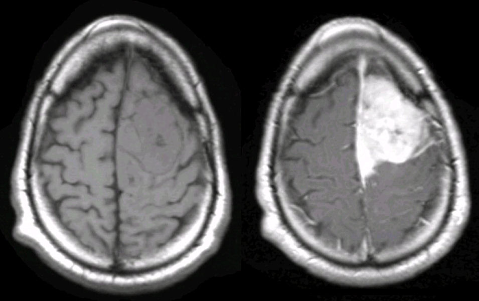
(Left) Axial MRI Scan T1 weighted; (Right) Axial MRI Scan T1 weighted with gadolinium. Note the large, well demarcated mass which is dural based and compressing the adjacent frontal lobe. The mass is isodense on T1 but strongly enhances with contrast. This is the typical appearance of a meningioma. Meningiomas are common tumors. They are typically benign histologically, and treatable with surgical resection if they are in an accessible location.
Last Update:
11/8/05
The Electronic Curriculum is copyrighted 1998, Case Western Reserve University
School of Medicine.